Figure 6.

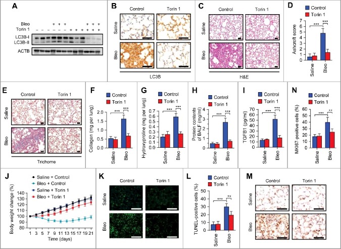
Pharmacological activation of TFEB by Torin 1 elevates autophagic flux and ameliorates pulmonary fibrosis in bleomycin-treated mice. Mice were injected intratracheally with bleomycin or saline, and treated with Torin 1 (20 mg/kg) every other day for 21 d (n = 5 per group). (A) Lung tissues were homogenized and immunoblotted for LC3B. ACTB, loading control. (B) Representative images of LC3B immunohistochemical staining of lung tissue sections. Scale bars: 50 μm. (C) Representative images of hematoxylin and eosin (H&E) staining of lung tissue sections. Scale bars: 50 μm. (D) Ashcroft fibrotic scores were determined by H&E staining of lung sections. (E) Representative images of Masson's trichrome staining of lung tissue sections. Scale bars: 50 μm. (F and G) Whole lung homogenates were analyzed to examine collagen contents using the Sircol assay (F) and hydroxyproline contents (G). (H and I) Total protein contents (H) and TGFB1 levels (I) were determined in bronchoalveolar lavage fluid (BALF). (J) Body weights were measured over time. (K) Representative images of TUNEL staining of apoptotic cells in lung tissue sections. Scale bars: 50 μm. (L) Quantification of apoptotic cells shown in (A). (M) Representative images of MKI67 immunohistochemical staining in lung tissue sections. Scale bars: 50 μm. (N) Percentage of MKI67-positive cells shown in (M). Data are means ± s.d. **P < 0.01, ***P < 0.001 (Student t test).
