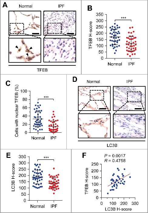
Figure 7.

Expressions of TFEB and LC3B are negatively associated with human pulmonary fibrosis. (A) Representative images of TFEB immunohistochemical staining in pulmonary fibrosis (PF) tissues or adjacent normal lung tissues from patients. Arrows represent lung epithelial cells with nuclear TFEB staining. Scale bars: 50 μm. (B and C) TFEB staining intensity (B) and percentage of nuclear TFEB-positive cells (C) of lung epithelial cells in 41 PF tissues and paired adjacent normal lung tissues from patients. (D) Representative images of LC3B immunohistochemical staining in pulmonary fibrosis (PF) tissues or adjacent normal lung tissues from patients. Scale bars: 50 μm. (E) LC3B staining intensity in PF tissues and paired adjacent normal lung tissues from patients (n = 41). (F) Association of the staining intensity of TFEB and LC3B in PF tissues (n = 41). Concordance was determined by Pearson correlation and linear regression. Data in (B, C and E) are means ± s.d. Results are representative of 3 independent experiments unless stated otherwise. ***P < 0.001 (Student t test).
