Figure 2.

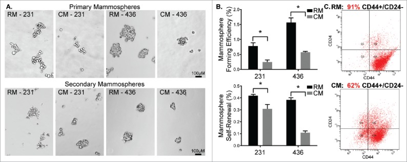
MDA-MB-231 and MDA-MB-436 cells treated with mESC CM display diminished CSC activity. MDA-MB-231 and MDA-MB-436 cells were pre-treated with RM or CM after which they were utilized in mammosphere assays. (A) Representative images of primary mammosphere formation and representation of the potential for self-renewal following passaging of primary mammospheres to form secondary mammospheres. (B) Quantification of primary mammosphere forming efficiency and secondary mammosphere self-renewal. Additionally, MDA-MB-231 cells exposed to RM and mESC CM were assessed for stem/progenitor cell surface markers using (C) flow cytometry to determine changes in the CD44+ / CD24− subpopulation of cells, indicative of a stem/progenitor phenotype. Statistical significance indicated by * for p<0.05. Scale bars are 100 μm.
