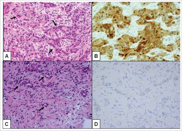
Figure 1.

Histologic review of the second biopsy showed high grade pancreatic ductal adenocarcinoma (PDA), metastatic to the liver throughout the tissue cores (original magnification 400X). (A) Areas of undifferentiated tumor cells containing markedly pleomorphic nuclei were arranged in highly cellular nests and clusters of malignant cells (arrows) within a relatively scarce fibrotic stroma. (B) These undifferentiated tumor cells were positive by immunohistochemistry for IDH1 (R132H) mutation. (C) Distinct areas of the tumor biopsy demonstrated less tumor pleomorphism that was more characteristic of conventionally tubular morphology (arrows). (D) In contrast to high grade foci, the well-differentiated foci were negative by immunohistochemical stain for IDH1 (R132H) mutation, consistent with IDH1 wild type status of tumor cells in these areas.
