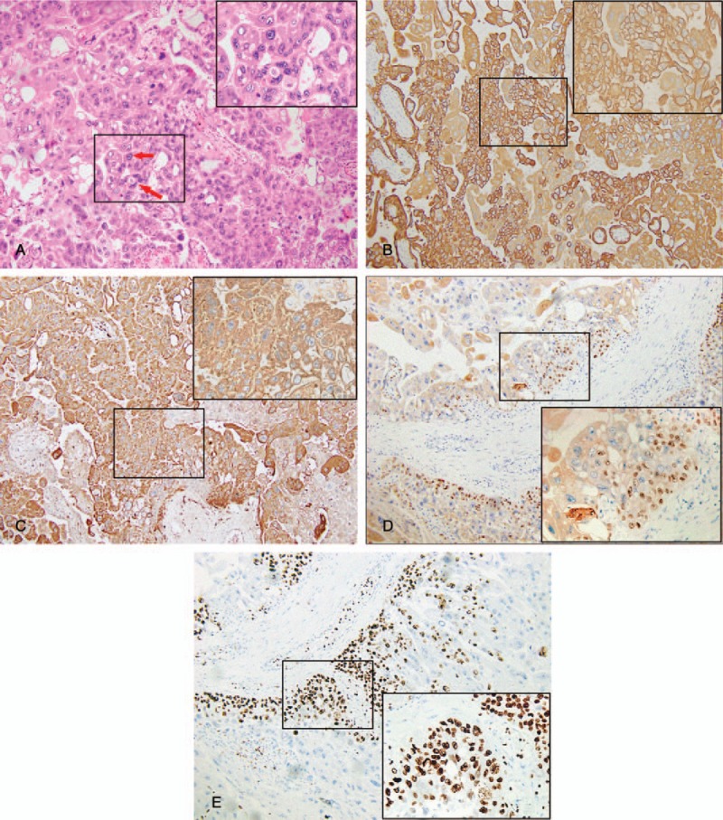
Figure 1.

(A) Representative images of hematoxylin and eosin stain. The top right corner (400× of the image shows an enlarged image in A (100×). The arrows indicate marked cytological atypia, with variably sized nucleus. (B) Representative images of immunohistochemistry staining for CK. The top right corner (400×) of the image shows an enlarged image in B (100×). And this image shows positive expression of CK in choriocarcinoma cells. (C) Representative images of immunohistochemistry staining for HCG. The top right corner (400×) of the image shows an enlarged image in C (100×). And this image shows positive expression of HCG in choriocarcinoma cells. (D) Representative images of immunohistochemistry staining for P63. The bottom right corner (400×) of the image shows an enlarged image in D (100×). And this image shows positive expression of P63 in choriocarcinoma cells. (E) Representative images of immunohistochemistry staining for Ki-67. The bottom right corner (400×) of the image shows an enlarged image in E (100×). And this image shows positive expression of Ki-63 in choriocarcinoma cells. CK = cytokeratin, HCG = beta-human chorionic.
