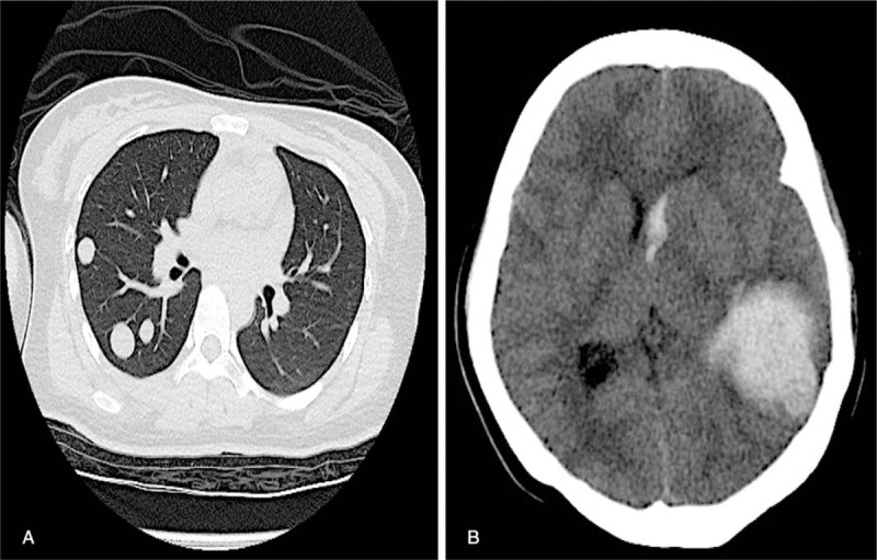
Figure 2.

(A) Chest computed tomography (CT) scan carried out at 95 days postdelivery showing several metastatic nodules in the bilateral pulmonary tissue. (B) The cerebral CT carried out at 100 days postdelivery showing cerebral hemorrhage (left temporal lobe) and a midline shift of the brain.
