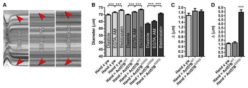
Figure 4. Excessive Ca2+-Independent Actomyosin Associations during Diastole Promote Enhanced Myocyte Shortening and Incomplete Relaxation of Hand > Act57BA295S Cardiomyocytes.
(A) M modes generated from the same region of a three-week-old Hand > Act57BWT heart reveal graded responses of cardiac diameters following exposure to distinct small-molecule compounds. Red arrowheads indicate the position of the heart wall edges during diastole, upon incubation with EGTA/EGTA,AM, and finally, following the addition of blebbistatin. Incubation with EGTA/EGTA,AM resulted in complete cessation of wall motion. Relative to the diameter during diastole, each treatment induced a slight increase in diameter across the heart tube.
(B) Significant, incremental increases in cardiac diameters were verified for all genotypes following extra- and intracellular Ca2+ chelation and upon blebbistatin incubation.
(C) The average change in diameter across the heart wall in response to EGTA/EGTA,AM was similar among all lines.
(D) Blebbistatin treatment prompted a significantly greater response across the wall of Hand > Act57BA295S hearts relative to that observed for Hand × yw and Hand > Act57BWT hearts.
Data are presented as mean ± SEM (n = 21). ***p ≤ 0.001. See also Figure S3.

