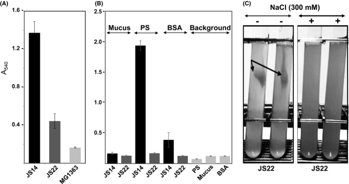
Figure 3.

Biofilm formation and adherence of JS14 and JS22 to different surfaces. (A) Biofilm formation of JS14, JS22 and the non‐biofilm‐forming L. lactis control. After 3 days of incubation at 30°C, the washed biofilms were stained with crystal violet and quantified using an ELISA reader. The error bar indicates standard deviation (SD) for three biological and eight technical replicates. (B) Adhesion of JS14 and JS22 to porcine mucus, BSA and PolySorp (hydrophobic surface). Adherence of the cells was quantified using the crystal violet staining method as described above. The error bars indicate SD for three independent tests, each with sixteen technical replicates. (C) Growth of JS22 with or without NaCl. The test tubes represent JS22 cells cultured in PPA with and without 300 mM NaCl for 72 h under microaerophilic conditions at 30°C. Arrows indicate the formed clumps.
