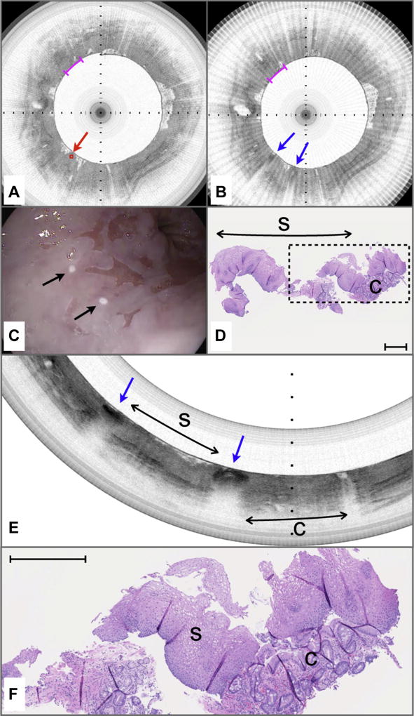
Figure 6.
Volumetric laser endomicroscopy (VLE)–guided biopsy of where the pre-marking and post-marking diagnoses in the study aspect ratio did not correspond to the majority histopathologic diagnosis. A, An area containing optical coherence tomography (OCT) features of columnar-lined esophagus (c) is identified as an intent-to-biopsy region (center of target region delineated by red diamond and arrow). B, After the marking laser is activated, the superficially cauterized mucosa can be seen as darkened regions in this post-marking VLE image (blue arrows). The region between the marks also was diagnosed as columnar-lined esophagus by the blinded OCT reviewer when it was assessed in the study aspect ratio as displayed. C, Videoendoscopy performed after the balloon-centering catheter was withdrawn shows the 2 laser cautery marks (black arrows) that are apparently on squamous mucosa. D, Histology from the biopsy specimen obtained at the marks shows a predominance of squamous epithelium with a small portion of columnar epithelium with intestinal metaplasia. E, The post-marking image displayed at the correct full aspect ratio and filtered to remove the radial striping artifact seen in B. This image demonstrates that the area between the marks has a layered appearance that is consistent with squamous mucosa (s). The rightmost mark borders tissue with OCT features of columnar mucosa (C). F, An expanded view of the histology in D shows squamous mucosa with a fragment of columnar-lined esophagus with intestinal metaplasia. Black tick marks in A and B, 350 µm in depth. Magenta scale bars in A and B, 3 mm along the circumference. Scale bar in D, 500 µm. Tick marks in E, 1 mm.

