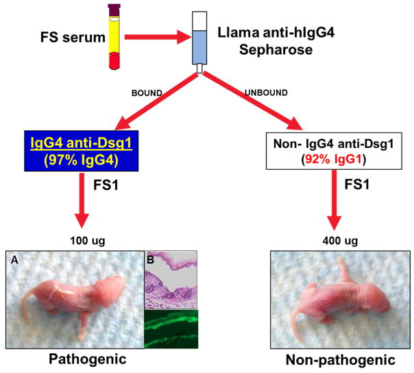
Figure 1. FS IgG4 autoantibodies are pathogenic.
The IgG4 fraction from FS1 serum (from patient #1 in the set of 20 tested, FS1 is used throughout the paper) was purified on a llama anti-human IgG4 Sepharose affinity column. The bound fraction contained 97% pure IgG4. The unbound fraction contained non-IgG4 subclasses with IgG1 representing the majority (92%). The IgG4 and non-IgG4 fractions were dialyzed, concentrated and injected subcutaneously into neonatal mice. A. Mice injected with FS1 IgG4 (left) developed extensive blistering, shown as fine wrinkling of the epidermis (induced by slight friction or pinching, the so-called Nikolsky’s sign). B. On histological examination large sheets of epidermis were separated from the naked dermis (x200) and direct immunofluorescence shows bound IgG4 to the roof of the split (x100). Mice receiving a higher dose of non-IgG4 fraction showed no disease (right).

