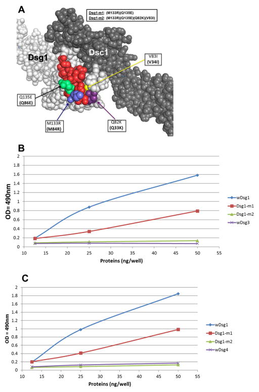
Figure 7. Identification of key amino acid residues for FS IgG4 binding.
(A)The locations of the mutated residues (blue, lime green, yellow and dark purple) in the EC1 epitope (red) are shown on the homology model of the Dsg1/Dsc1 heterodimer (light (Dsg1) and dark (Dsc1) gray). (B) Affinity-purified FS1 IgG4 antibodies on the llama anti-human IgG4 and Dsg1-EC1/Dsg3 bb columns were tested against wDsg1 (blue), Dsg1-m1 (red), Dsg1-m2 (green) and wDsg3 (purple) by ELISA. A stock of IgG4 (0.3 mg/ml) from each patient at a dilution of 1:5,000 was tested on microtiter wells coated with different amounts of the recombinant protein (from 12.5 ng/well to 50 ng/well). Peroxidase-labeled mouse anti-human IgG4 at a dilution of 1:2,000 was used as an indicator. The results are expressed in OD490nm units. (C) Affinity-purified FS1 IgG4 antibodies on the llama anti-human IgG4 and Dsg1-EC1/Dsg4 bb columns were tested against wDsg1 (blue), Dsg1-m1 (red), Dsg1-m3 (green) and wDsg4 (purple) by ELISA. A stock of IgG4 (0.3 mg/ml) from each patient at a dilution of 1:5,000 was tested on microtiter wells coated with different amounts of the recombinant protein (from 12.5 ng/well to 50 ng/well). Peroxidase-labeled mouse anti-human IgG4 at a dilution of 1:2,000 was used as an indicator. The results are expressed in OD490nm units.

