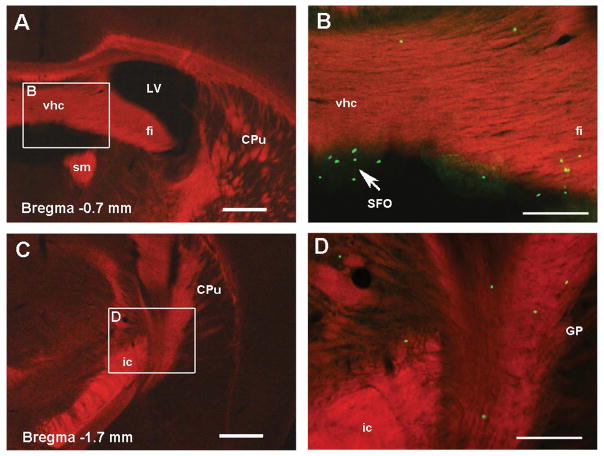
Fig 5. GFP+ cells in the white matter of adolescent Recon Rag2−/− mice.
Fluorescent microscopy images of coronal brain sections of adolescent Rag2−/− mice neonatally reconstituted with GFP+ lymphoid cells. Panels A and C show white matter structures labeled with FluoroMyelin Red (100X magnification). Higher magnification (200X) of the delineated regions are merged with the green channel in Panels B and D to show the localization of GFP+ cells in the white matter and circumventricular organs (arrow). Bregma coordinates correspond to the adult mouse atlas from Franklin and Paxinos, Third ed. 2007. CPu: caudate putamen, fi: fimbria of the hippocampus, GP: globus pallidus, ic: internal capsule, LV: lateral ventricle, SFO: subfornical organ, sm: stria medullaris thalamus, vhc: ventral hippocampal commissure. Scale bar in A, C: 400 μm, B, D: 200 μm.

