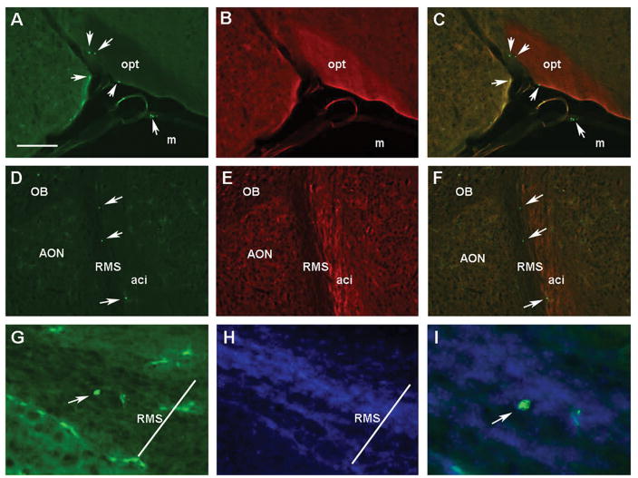
Fig. 6. GFP+ lymphocytes in the brain of adult Recon Rag2−/− mice.
Fluorescent microscopy images of GFP+ cells (green) and FluoroMyelin Red stained coronal (A–C) and horizontal (D–F) brain sections of adult Rag2−/− mice neonatally reconstituted with GFP+ lymphoid cells. Panels A–C show a coronal section showing the optic tract (opt) where GFP+ cells can be found the meninges (m), as well as within the brain. Images show the green (A), red (B) and C) merged channels, respectively. Panels D–I correspond to horizontal sections shown at the level of the anterior olfactory nucleus (AON); D: GFP, E: FluoroMyelin Red, F: merged. GFP+ cells are localized in the rostral migratory stream (RMS) next to the anterior commissure, intrabulbar (aci). G) A GFP+ cell is seen inside the boundaries of the RMS at higher magnification (200X); H) Hoechst nuclear counterstain. I) A merged image showing the GFP+ cell at 400X magnification. Arrows point to identified GFP+ cells (all panels). Scale bar in A = 250 μm for panels A–F. Transverse bar in G & H = 70 μm denoting the approximated width of the RMS. OB: Olfactory bulbs.

