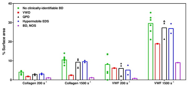
Figure 5.
Percentage of flow channel covered with platelets and/or and platelet aggregates for each study participant at shear rates of 200 s−1 and 1500 s−1 on VWF and collagen surfaces. The participant symbols are color-coded by diagnosis: green indicates no clinically-identifiable BD; red indicates VWD; black indicates QPD; blue indicates hypermobile EDS; and purple indicates BD, NOS. Participant 5 also has a thrombocytopenia diagnosis and this is indicated by open symbols. This assay was not performed for participant 10.

