Figure 2.
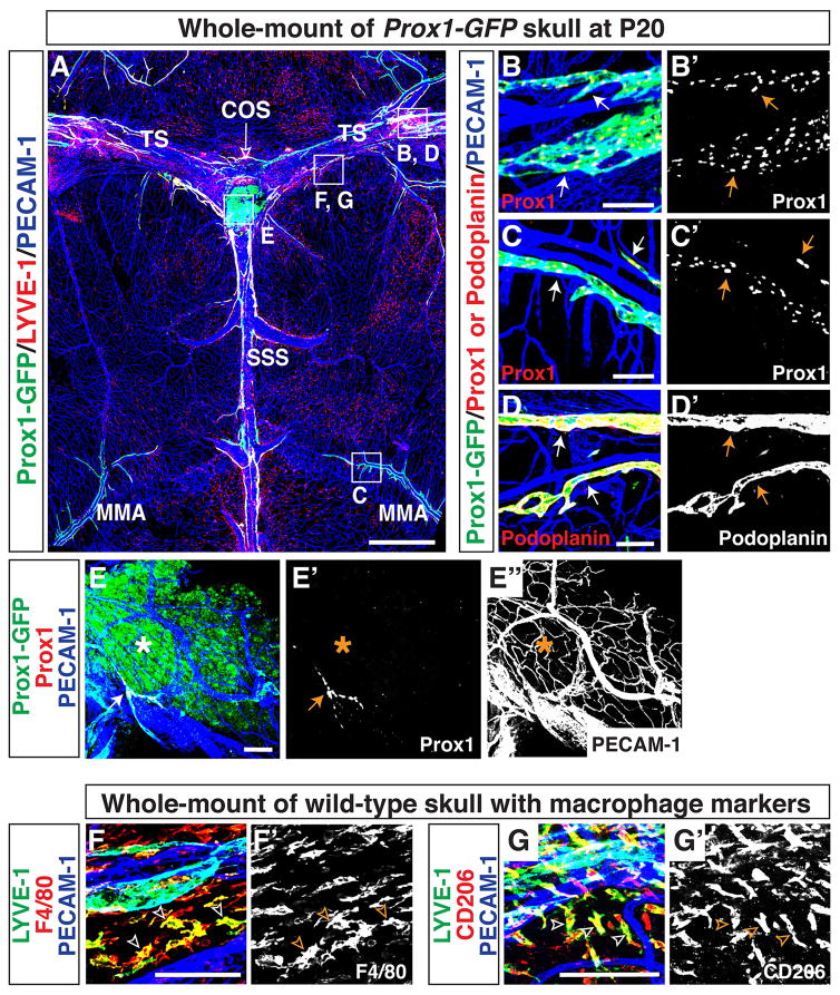
Lymphatic vessels in the postnatal brain meninges express well-established lymphatic markers. (A) Lymphatic vessel patterning at postnatal day (P)20 is similar to the patterning in adult. In addition to lining the SSS, TS, COS (open arrow), and MMA, lymphatic vessels extend along additional smaller blood vessels in the meninges. The boxed regions in (A) are magnified in (B–G), with staining using different antibodies. (B-B′) Lymphatics along the TS are positive for both Prox1-GFP and Prox1 proteins (arrows). (C-C′) Lymphatics along the MMA are positive for both Prox1-GFP and Prox1 proteins (arrows). (D-D′) Lymphatic vessels along the TS are positive for lymphatic marker Podoplanin (arrows). (E-E″) The Prox1-GFP-positive area (asterisk) at the COS (open arrow) is not stained with anti-Prox1 antibody, indicating that this is not a real lymphatic region. Note that a small-diameter Prox1/PECAM-1-double positive lymphatic vessel (E and E′, arrow) and a PECAM-1-positive blood vascular network (E and E″) are observed at the COS. (E–F′) LYVE-1-positive cells in the meninges are also positive for the macrophage markers F4/80 (F-F′, open arrowheads) and CD206 (G-G′, open arrowheads) indicating that LYVE-1 single-positive cells in (A) are a subset of tissue macrophages. Scale = 1mm in (A) and 100 μm in (B–G, B′–G′).

