
Fig. 5.
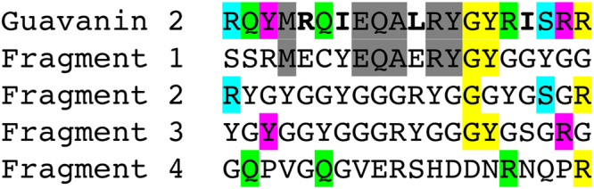
Guavanin 2 and its ancestors. Guavanin 2 and the Pg-AMP1 fragments were aligned without including gaps to demonstrate the guavanin 2 inherited residues and mutations. The residues inherited from each the fragments are highlighted in gray (fragment 1—α-helix propensity), cyan (fragment 2—net charge), magenta (fragment 3—hydrophobicity), light green (fragment 4—hydrophobic moment), and yellow (two or more fragments); and the mutated residues are in bold face