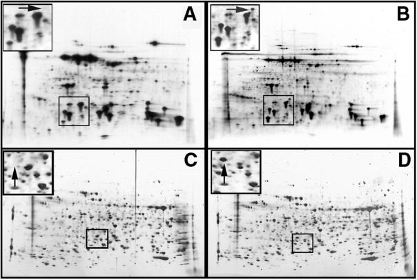
Figure 5.
Autoradiographs (A and B) and silver stains (C and D) of 2D-PAGE electrophoresis separation of proteins from AtTS02 (A and C) and RLD (B and D) seedlings following pre-incubation at 38°C for 4 h to induce acquired thermotolerance. Insets represent magnified sections of the gels showing the absence of a 27-kD protein in the AtTS02 seedlings (A and C, arrow) compared with the RLD seedlings (B and D, arrow).

