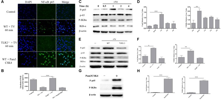
FIGURE 4.
Trichomonas vaginalis induces changes of subcellular localization of NF-κB p65 via TLR2. Confocal microscopy analysis revealed the effects of T. vaginalis or Pam3CSK4 on the translocation of NF-κB p65 from the cytoplasm to the nucleus in WT macrophages after the incubation with T. vaginalis for 1 h (A). The percentage of colocalization of NF-κB with the nuclear signal under the different treatments (B). WT mouse macrophages were incubated with T. vaginalis for different times (0–4 h), cell lysates were used for western blot analysis (C). WT and TLR2-/- mouse macrophages were incubated with T. vaginalis for 1 h, phosphorylation of NF-κB p65 and IκBα was detected by western blot (E). Phosphorylation of NF-κB p65 in Pam3CSK4 treated macrophages (G). Relative Gray analysis of western blot (D,F,H).

