Fig. 6.
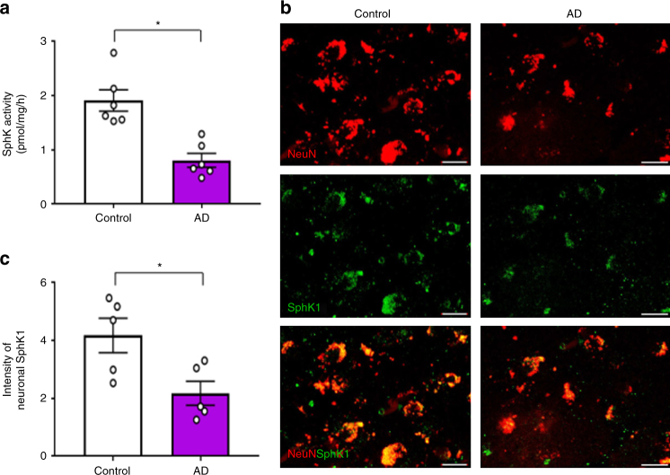
SphK1 is decreased in AD patient brains and neurons. a Characterization of SphK activity in cortex brain samples from AD and control human subjects (n = 6 per group). b Representative immunofluorescence images of cortex brain samples from AD and control human subjects showing SphK1 (green) merged with neuron (NeuN, red). Scale bars, 20 μm. c Quantification of neuronal SphK1 (n = 5 per group). a, c Student’s t test. *P < 0.05. All error bars indicate s.e.m.

