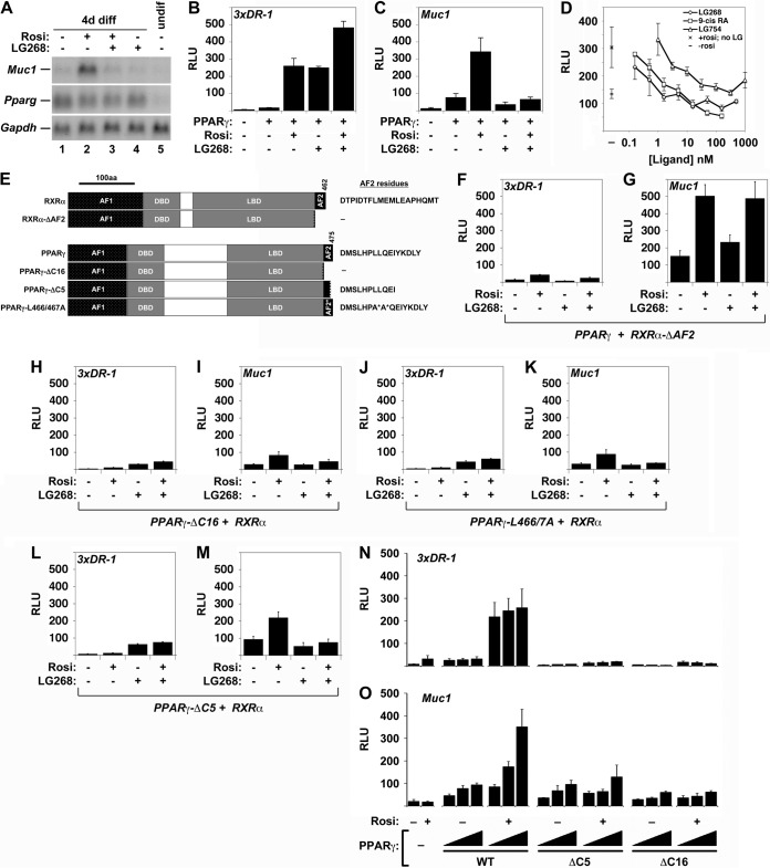
FIG 1.
Effects of rexinoids and RXRα and PPARγ AF2 domain mutations on Muc1 promoter activity. (A) Northern blot analysis of Muc1, Pparg, and Gapdh (normalization control) in cultures of the TSC line GFP-Trf differentiated for 4 days (4d diff) without treatment or in the presence of 1 μM Rosi, 1 μM LG268, or both, as well as an undifferentiated culture (undif), as indicated. (B) Relative light units (RLU) in extracts of CV1 cells transiently transfected with pCMX-βGAL, a 3xDR1-luciferase (3xDR1-luc) construct, and RXRα alone versus RXRα and PPARγ with the indicated combinations of Rosi and LG268, normalized to β-galactosidase activity. (C) Same as described for panel B, with a Muc1-luc construct instead of 3xDR1-luc. (D) Normalized RLU in CV1 cells transfected with pCMX-βGAL, Muc1-luc, RXRα, and PPARγ and treated with 1 μM Rosi and incremental concentrations of the distinct rexinoids LG268, 9-cis retinoic acid, and LG754, from 0.3 nM to 1 μM. Left, mean basal RLU ± standard error (SE) without RXR ligands: bottom, no Rosi (–); top, 1 μM Rosi (×). Right, mean RLU ± SE of experimental data. (E) Summary of RXRα and PPARγ AF2 domain mutants used. Exact AF2 residues present in each species are shown. (F to M) Normalized RLU in CV1 cells transfected with pCMX-βGAL, either 3xDR1-luc (F, H, J, L) or Muc1-luc (G, I, K, M), and the indicated RXRα or PPARγ combinations: full-length (FL)-PPARγ + RXRα-ΔAF2 (F, G), PPARγ-ΔC16 + FL-RXRα (H, I), PPARγ-L466A/L467A + FL-RXRα (J, K), and PPARγ-ΔC5 + WT RXRα (L, M). The results shown in panels B, C, and F to M are part of a transfection series performed side by side and are directly comparable. (N, O) Dose-dependent effects of FL-PPARγ, PPARγ-ΔC5, and PPARγ-ΔC16 on the Muc1 and DR1 reporters. Normalized RLU in CV1 cells transfected with pCMX-βGAL, RXRα, and 3xDR1-luc (N) or Muc1-luc (O) and one to three quanta of the indicated PPARγ AF2 domain configurations (WT, ΔC5, and ΔC16), incubated in the absence or presence of 1 μM Rosi, as indicated; a filler plasmid (pCMX-GAL4N) was used to equalize the DNA concentration in reaction mixtures containing less than three quanta of the PPARγ variants. Bars and error bars show mean values and SE.

