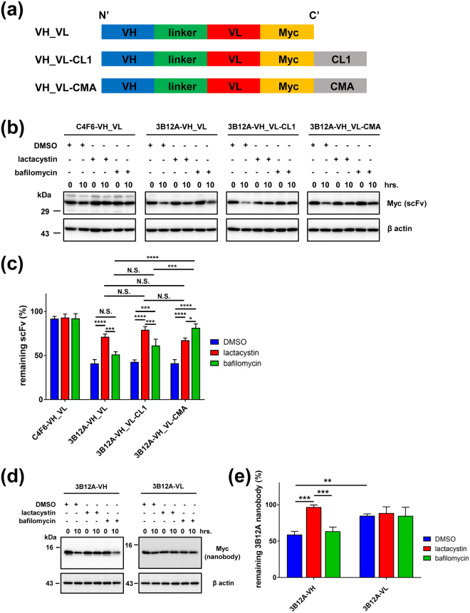
Figure 3.
3B12A scFv-CMA is degraded by both proteasome and autophagy proteolytic pathways. (a) Illustrative domain profiles of 3B12A scFv for VH_VL-Myc with or without proteolytic signals including CL1 for proteasomes and chaperone-mediated autophagy (CMA) for autophagosomes. (b) Protein degradation chase assay of C4F6 scFv (C4F6-VH_VL), 3B12A scFv (VH_VL), 3B12A scFv-CL1 (VH_VL-CL1) and 3B12A scFv-CMA (VH_VL-CMA) in HEK293A cells. (c) Quantitative analysis of (b) Each data point was obtained by normalisation to actin. Differences were evaluated by one-way ANOVA (mean ± SD from three independent experiments; *p < 0.05, ***p < 0.005 and ****p < 0.001). N.S. indicates not significant. ‘remaining scFv (%)’ indicates ‘the % signal compared with time 0’. (d) Protein degradation chase assay of 3B12A-VH and 3B12A-VL in HEK293A cells. (e) Quantified analysis of (d) Each data point was obtained by normalisation to actin. Differences were evaluated by one-way ANOVA (mean ± SD from three independent experiments; **p < 0.01 and ***p < 0.005).

