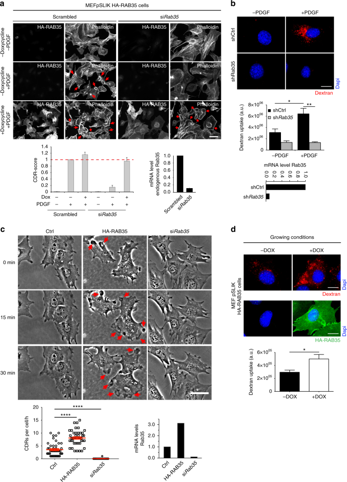
Fig. 2.
RAB35 impairs CDRs formation. a An siRNA-resistant human RAB35 rescues CDRs formation. Doxycycline-treated pSLIK-HA-RAB35-human infected MEFs silenced for endogenous RAB35 were serum starved for 2 h, and stimulated with PDGF for 10′. Samples were co-stained with FITC-Phalloidin and the anti-HA antibody to detect F-actin and human HA-RAB35, respectively. Red arrows indicate CDRs. Scale bar, 50 µm. CDR score was calculated by normalizing the number of CDR-positive cells per each condition against the scrambled doxycycline-untreated, PDGF-stimulated sample, used as a control. Data are the mean ± SD (n > 100 cells/condition, three independent experiments). The silencing of endogenous RAB35 was verified by qRTPCR. b Silencing of RAB35 impairs macropinocytosis. MEF control (shCtrl) and Rab35-downregulated (shRab35) cells were serum starved for 2 h and incubated (+) or not (−) for 1 h with PDGF and tetramethylrhodamine-dextran. Upon fixation, cells were processed for epifluorescence to identify nuclei (blue) and TMR-dextran-positive macropinosomes (red), respectively. Scale bar, 20 µm. Bottom graph, dextran uptake was quantified by determining the total cell fluorescence/cell (see Methods) expressed as A.U. Data are the mean ± SD (n = 40 cells/condition, three independent experiments). **p < 0.01; *p < 0.05. The downregulation of RAB35 was verified by qRTPCR. c RAB35 is sufficient to induce the spontaneous formation of CDRs. MEF control (Ctrl), HA-RAB35-expressing (HA-RAB35) and Rab35-silenced (siRab35) cells were monitored for 1 h by time-lapse in the absence of PDGF. Still phase contrast images from time-lapse sequence (Supplementary Movie 2) are shown. Red arrows indicate CDRs. Scale bar, 50 µm. The number of CDRs/cell formed in 1 h is expressed as mean ± SEM (n = 60 cells/condition, three independent experiments). ****p < 0.0001. RAB35 mRNA levels were determined by qRTPCR. d RAB35 expression promotes macropinocytosis in the absence of GFs. Doxycycline-treated pSLIK-HA-RAB35-human-infected MEFs were incubated for 1 h with TMR-dextran. Cells were processed for epifluorescence to visualize nuclei (blue), TMR-dextran-positive macropinosomes (red) and the ectopic expression of HA-RAB35 protein (green). Quantification of dextran uptake performed as in b. The total fluorescence/cell was expressed as A.U. Scale bar, 20 µm. Data are the mean ± SD (n = 40 cells/condition, three independent experiments). *p < 0.05. p-values are from paired Student’s t-test

