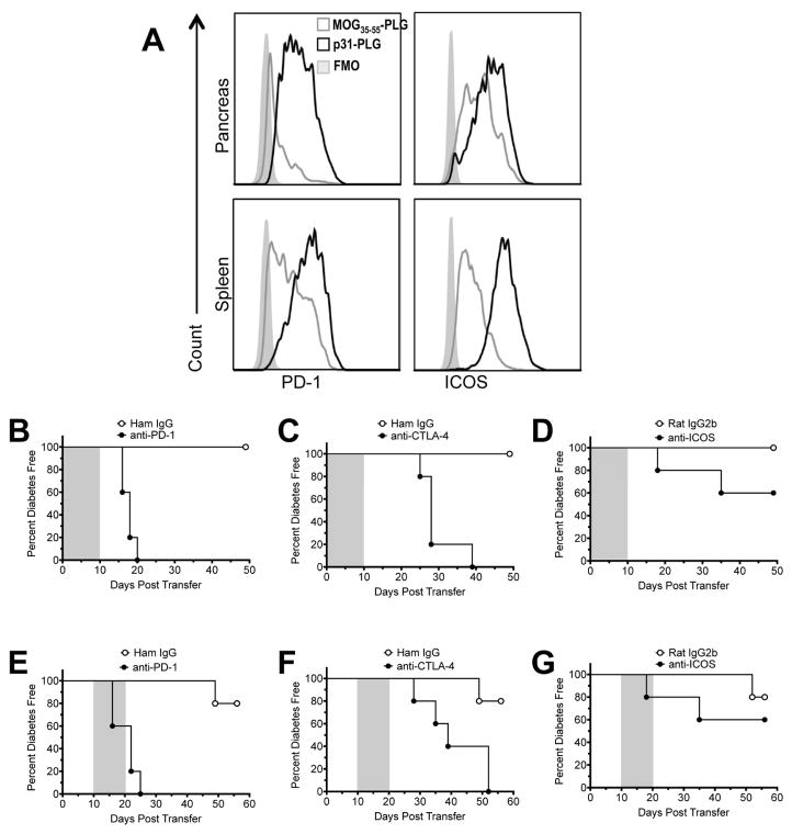
Fig. 5. Co-inhibitory signals are required for both induction and maintenance of Ag-PLG-induced tolerance.
(A) NOD.SCID recipients of 5×10⁶ p31 activated BDC2.5 T cells were tolerized with either MOG35–55-PLG/PEMA (N=3) or p31-PLG/PEMA (N=3) and expression of PD-1 and ICOS on T cells in the pancreas and SP was determined by FACs analysis on day +5. (B–G) NOD.SCID recipients of p31 activated BDC2.5 T cells were tolerized with p31-PLG/PEMA within 2 hours of cell transfer. Mice (N=5 per group) were then treated with anti-PD-1 (500μg first treatment, 250μg following treatments), anti-CTLA-4 (500μg), or anti-ICOS (500μg) blocking antibodies administered daily on days 0–10 (grey shading - B–D) or on days 10–20 (grey shading - E–G) post transfer and tolerization as indicated by the shaded area. Blood glucose levels were monitored weekly to determine diabetes incidence. Data is representative of two separate experiments.

