Figure 6.
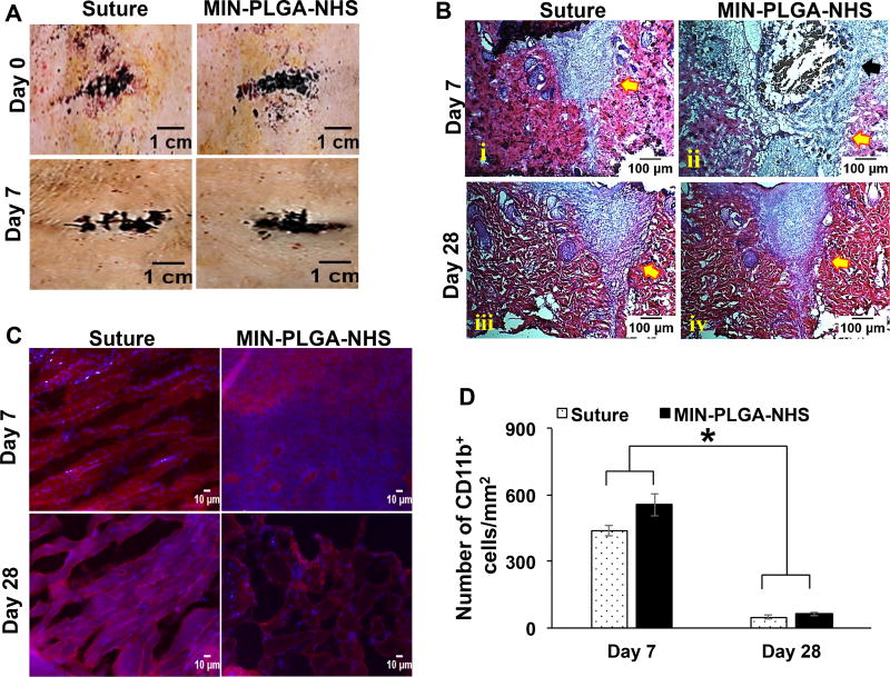
In vivo biocompatibility of mussel-inspired nanocomposites with PLGA-NHS nanoparticles (MIN-PLGA-NHS) (A) Micrographs of areas sealed by PLGA-NHS nanocomposites and the ones sutured on the back of Sprague-Dawley rats at Day 0 and Day 7 (B) H&E staining of wound areas that were sutured and adhered with MIN-PLGA-NHS at day 7 and day 28. (C) Immunohistochemistry staining of macrophages in tissue sections. (D) Quantification of number of cd11b+ cells per millimeter square area near the tissues adhered with MIN-PLGA-NHS and the suture.

