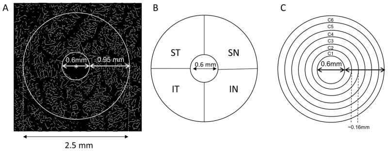
Figure 2. Partitions of the microvascular network.
The center (marked as red asterisk*) of the foveal avascular zone (FAZ) was detected in the skeletonized microvasculature image (A) and the center of the FAZ was used to partition the quadrantal and annular zones. The annulus from 0.6 mm to 2.5 mm in diameter was defined as the annular zone with a width of 0.95 mm after removing the avascular zone (diameter = 0.6 mm) centered on the fovea (A). Using the hemispheric partition, the annular zone was then partitioned into four quadrantal sectors (B), named the superior temporal (ST), inferior temporal (IT), superior nasal (SN), and inferior nasal (IN). To analyze the changes in the thin annular zones from the center to the periphery, the annular zone (A) was partitioned into 6 thin annuli named C1 to C6 (D) with a width of ~0.16 mm.

