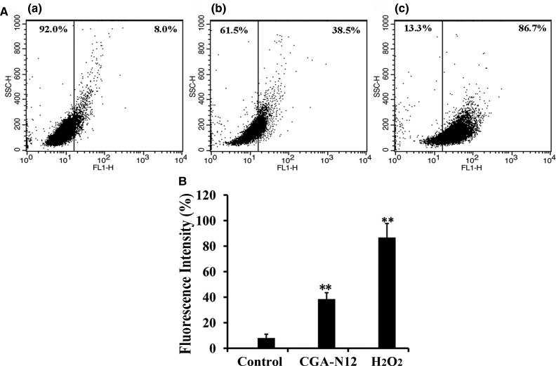
Figure 1. Detection of the effect of CGA-N12 on intracellular ROS accumulation.
(A) Approximately 1 × 10⁶ C. tropicalis cells were incubated with 75 µM CGA-N12 or 10 mM H2O2 for 10 h at 28°C. Intracellular ROS levels were detected by flow cytometry using dihydrorhodamine-123. Increases in fluorescence intensity indicate higher ROS levels. (a) Control cells that did not undergo CGA-N12 treatment; (b) cells exposed to 75 µM CGA-N12; (c) cells exposed to 10 mM H2O2. (B) Data represent the mean ± standard deviation for three independent experiments. Statistical significance was determined by Student's t-test. ** indicates statistical significant difference (P-values <0.01).

