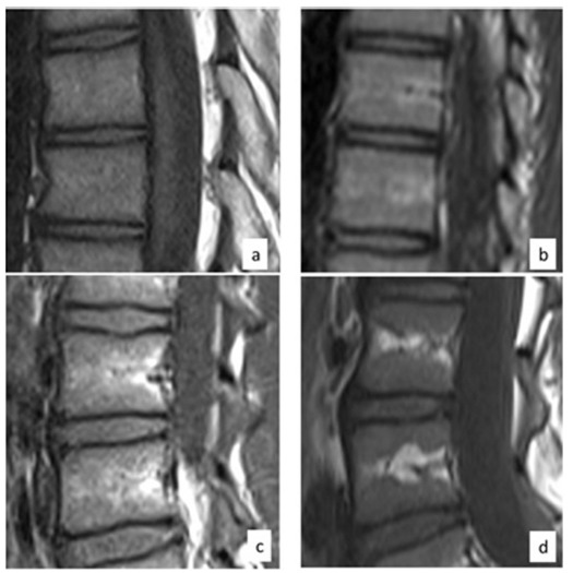
Fig. 1.

Type 1: overall T1-weighted signal increased compared with disc signal. No focal areas of increased T1-weighted signal, consistent with focal fatty change, are present (a). Type 2: linear areas of increase T1- weighted signal above and below the basivertebral vein, reflecting linear focal fatty change (b). Type 3: larger areas of increased T1-weighted signal reflecting increased areas of fatty change, either by thickening of the linear areas along the basivertebral vein (c) or wedge shape areas of increased T1-weighted signal along the peripheral central portion of the vertebral bodies (d).
