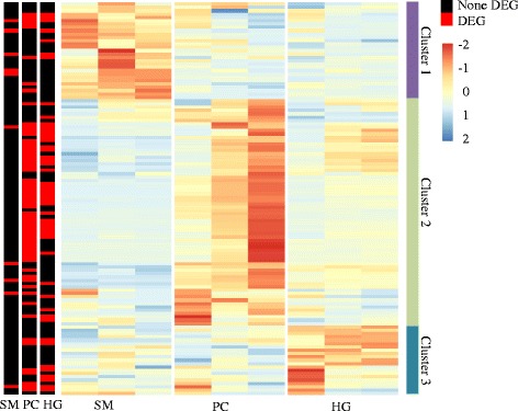
Fig. 1.

Expression of genes in phospholipid and lipoprotein synthesis pathways between different intestinal regions of salmon. For each tissue, the three columns represent 0.16 g, 2.5 g and 10 g samples from left to right. The color intensity is relative to the standard deviation from mean of TPM over developmental stages and tissues (row-scaled). Differential expressed genes (DEG, q < 0.05) between 0.16 g, 2.5 g and 10 g samples were annotated in three columns, which represent stomach (SM), pyloric caeca (PC) and hindgut (HG) respectively from left to right
