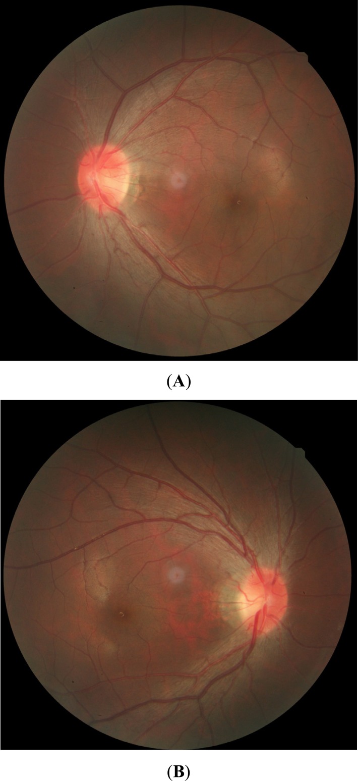
Fig. (1).
Fundus color images during the acute stage of LHON. (A, B) Swelling of the nerve fiber layer around the optic disc, circumpapillary telangiectatic microangiopathy, and presence of the optic nerve boundary can be observed. (For interpretation of the references to color in this figure legend, the reader is referred to the web version of this paper.)

