Fig. 1.

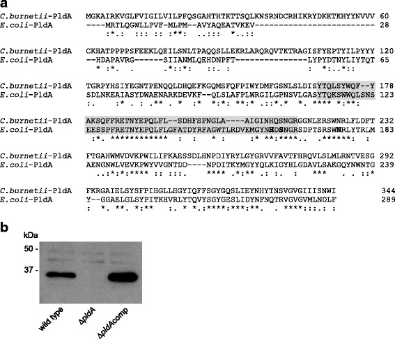
Generation of a C. burnetii pldA mutant. a Alignment of PldA from E. coli and C. burnetii. The grey region shows the consensus sequence motif YTQ-Xn-G-X2-H-X-SNG for PldA enzymes. Bold amino acids show known active site residues [15]. b Lysates from C. burnetii wild type, ΔpldA, and ΔpldAcomp strains after 6 days of growth in ACCM-2 (early SCV) were probed by immunoblotting with an anti-PldA polyclonal antibody
