Fig. 2.

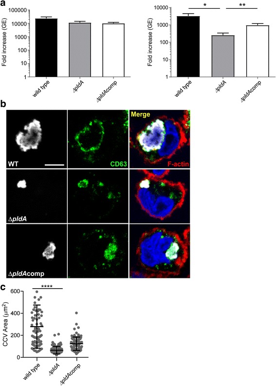
A ΔpldA mutant has impaired growth in THP-1 macrophages. a Growth of C. burnetii wild type, ΔpldA, and ΔpldAcomp strains in ACCM-2 (left panel) and THP-1 macrophages (right panel). Data represent fold increases in genome equivalents (GE) after 6 days of growth (early SCV) for 3 independent experiments performed in triplicate. Asterisks indicate a statistically significant difference (* = P < 0.05, ** = P < 0.01) as determined by the unpaired Student t test. b Immunofluorecence micrographs of C. burnetii wild type, ΔpldA, and ΔpldAcomp strains after 3 days of growth (LCV). Bacteria are colored white, the vacuole membrane green, the THP-1 cell border red, and the nucleus blue. c Size of Coxiella-containing vacuoles (CCV) at 3 days post-infection, Vacuole size was measured with Fiji and the data are representative of three independent experiments. Asterisks indicate a statistically significant difference. (**** = P < 0.0001). Scale bar, 5 μm
