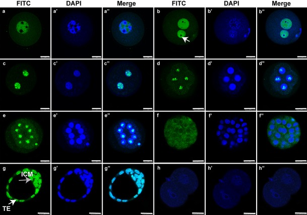
Fig. 5.
Immunofluorescence localization of SNAI3 during the germinal vesicle (GV) stage and early embryonic cleavages. Immunostaining with an anti-SNAI3 antibody (green) and DNA staining with 4′,6-diamidino-2-phenylindole dihydrochloride (blue). (a–a’’) GV. (b–b’’) One-cell stage (white arrow indicates the nucleolus). (c–c’’) Two-cell stage. (d–d’’) Four-cell stage. (e–e’’) Eight-cell stage. (f–f’’) Morula. (g–g’’) Blastocyst (ICM, inner cell mass, TE/white arrow, trophectoderm). (h–h’’) Negative control for SNAI3. Bars = 25 μm.

