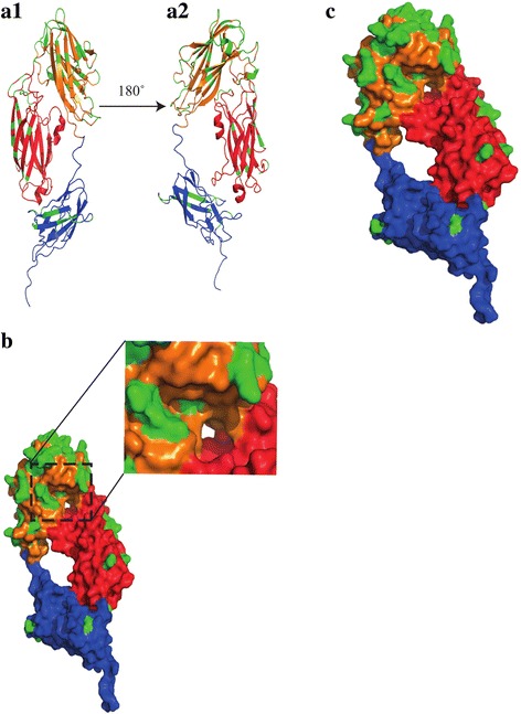
Fig. 5.

Structural characterization of the sdrD variants. 3D models were generated based on the N2-N3 and B1 subdomains of SdrD [15] in seven representative S. aureus strains. The subdomains are shown as N2 (red), N3 (orange) and B1 (Blue). Green colour coding indicates the variable amino acid residues within the seven sdrD variants (a) AI is ribbon diagram of N2-N3-B1 while A2 shows the diagram when reoriented 180 degrees. (b) Space filling diagram of N2-N3-B1. The grove between N2 and N3 is enlarged. (c) Space filling diagram of N2-N3-B1. The model shows the location of varied amino acids residues on the surface
