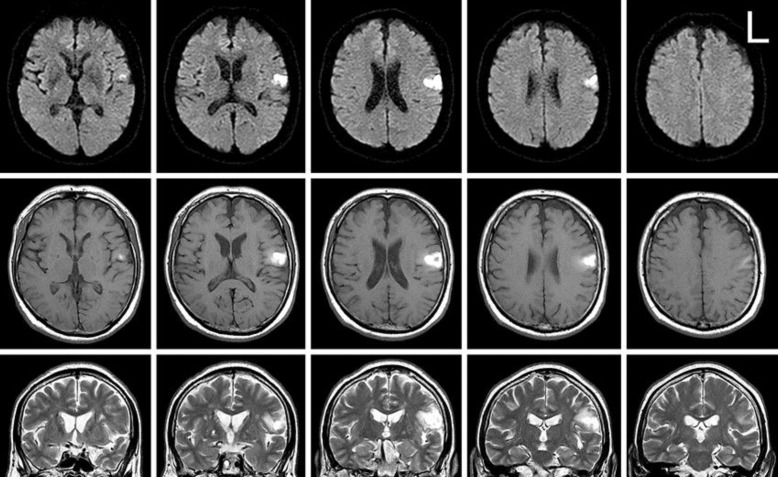
Fig. 1.
MR images at 13 days after onset. Diffusion-weighted axial (upper), T1-weighted axial (middle), and T2-weighted coronal (lower) images showed a hyperintense area, suggesting hemorrhage, under the opercular part of the left precentral gyrus (area 6) that extended upward under the postcentral gyrus. T2-weighted images also showed old lacunar infarctions in the basal ganglia and periventricular white matter bilaterally.

