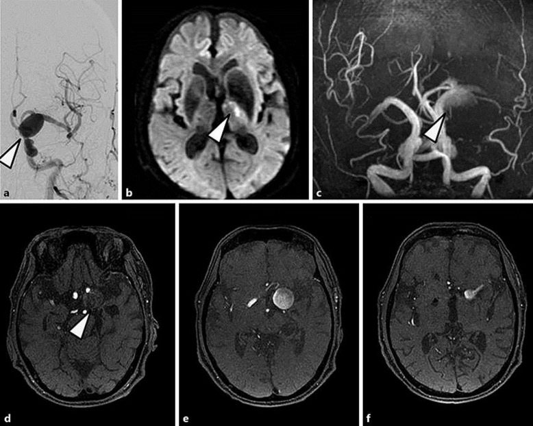
Fig. 1.
Digital subtraction angiography (a) obtained 5 years before admission. Magnetic resonance imaging (b–f) obtained on admission. a Left carotid angiography displays a large intracranial aneurysm (arrowhead; maximum diameter 21 mm) on the left internal carotid artery. b Diffusion-weighted imaging reveals a hyperintense signal on the left anterior choroidal artery territory (arrowhead). c–e Time-of-flight magnetic resonance angiography demonstrated poor visibility of the middle and anterior cerebral arteries and the inferior part of the giant aneurysm (maximum diameter 28 mm), suggesting aneurysm thrombosis (arrowheads).

