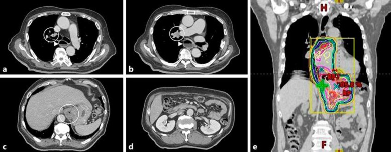
Fig. 2.
Computed tomography (CT) findings before radiotherapy. a Metastasis to the pretracheal lymph node at the level of the esophagus. b Right hilar lymphadenopathy at the level of the esophagus. c Gastric cardia (primary lesion). d No metastasis was visible in the para-aortic lymph node. e The radiotherapy dose distribution.

