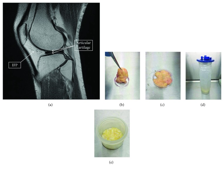
Figure 1.
(Modified and used with permission from Wiley under CC BL). Infrapatellar fat pad (IFP) location and harvested tissue. (a) Sagittal magnetic resonance imaging scan of the knee showing the relationship of the IFP (arrow) to the articular cartilage (double arrow). (b, c) Excised IFP from a patient undergoing knee arthroplasty (b) has the fat removed from the fibrous tissue (c). (d, e) The arthroscopically harvested fat pad (d) was separated from the irrigation fluid before enzymatic digestion (e).

