Figure 1.
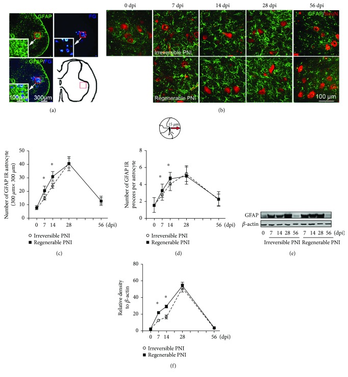
The pattern of astrocyte activation in the spinal ventral horn after sciatic nerve injury. (a) Representative cross sections of L5 spinal cord showing GFAP-positive astrocytes and FG retrograde-labeled motoneurons that are mainly localized in the spinal ventral horn. So, a schematic diagram indicating a 300 μm × 300 μm square area was defined for further morphometric analysis. (b) GFAP/NeuN double immunostaining showing the activated astrocytes (green) and motoneurons (red) in the area as (a) defined in different groups. (c) Quantitative analysis of the number of GFAP-positive astrocytes in each measured area. (d) The number of GFAP-positive processes (measured at 15 μm away from the soma) in each astrocyte. (e-f) Immunoblot analysis of GFAP expression in the L5 spinal ventral horn. ∗P < 0.05.

