Fig. 5.
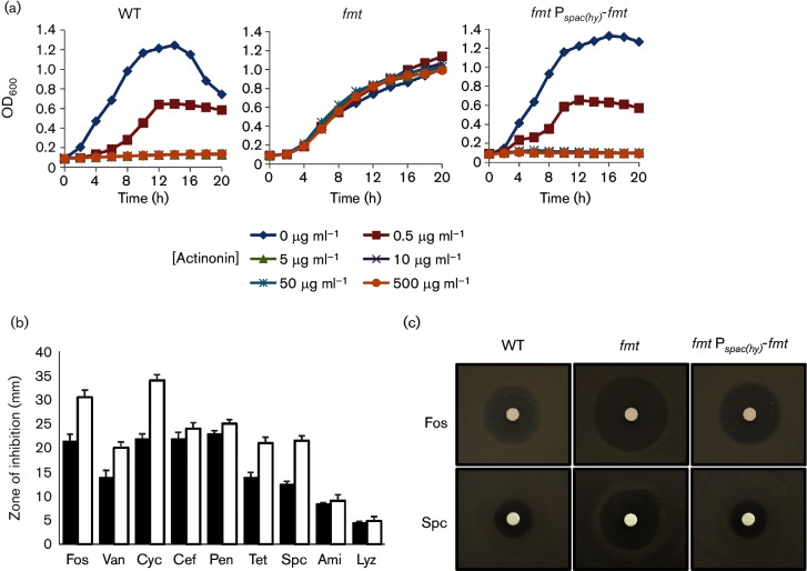
An fmt mutant displays altered antibiotic sensitivity. (a) Sensitivity of WT, isogenic fmt :: kan mutant (HB21006) and the fmt :: kan Pspac-fmt (HB21016) strains to actinonin as measured by growth curve assays. Actinonin concentrations used are noted below the graph. The data shown are representative of at least three independent experiments. (b) Sensitivity of WT (black bars) and an isogenic fmt :: kan mutant (HB21006, white bars) to antibiotic as monitored using a disc diffusion assay on LB plates. Antibiotics and cell envelope active agents used were fosfomycin (Fos, 250 µg), vancomycin (Van, 50 µg), d-cycloserine (Cyc, 1 mg), cefuroxime (Cef, 6 µg), penicillin G (Pen, 100 µg), tetracycline (Tet, 50 µg), spectinomycin (Spc, 0.5 mg), amitriptyline (Ami, 250 µg) and lysozyme (Lyz, 200 µg), The mean±se from at least three biological replicates is reported. (c) Representative photographs (from at least six replicates) of a disc diffusion assay with WT or an fmt mutant (HB21006) and an fmt Pspac(hy)-fmt complemented strain (HB21016) cell on LB plates. The discs were spotted with fosfomycin (250 µg) or spectinomycin (0.5 mg) as indicated.

