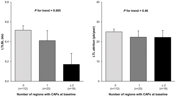
Figure 2. Baseline leukocyte telomere length (left) and leukocyte telomere attrition during the follow-up period (right) versus the number of regions with carotid atherosclerotic plaques at baseline.
Values are mean ± SEM. BL, baseline; CAP, carotid atherosclerotic plaque; LTL, leukocyte telomere length; kb, kilo base pairs; bp, base pair.
BL LTL is adjusted to age and sex; LTL attrition is adjusted to age, sex and baseline value of LTL.

