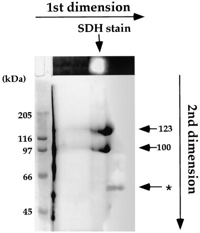
Figure 2.
Two-dimensional separation the purified soybean LKR/SDH. The purified fraction followed the Blue Sepharose column (Fig. 1f) was separated on a first-dimensional native gel and the gel was stained for SDH activity (top gel). The first-dimensional gel was separated on a second-dimensional SDS gel and the gel was stained with Coomassie Blue R-250. The positions of the 100-, 123-, and 60-kD (asterisk) polypeptides in the second-dimensional gel are indicated by arrows on the right. Sizes of protein markers are indicated on the left.

