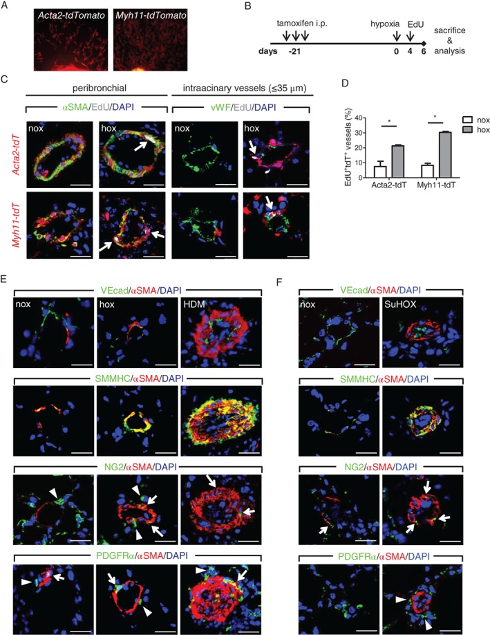
Figure 5.

In vivo and ex vivo expansion of resident vascular smooth muscle cells. (A) Outgrowth of tdTomato‐labeled SMCs from main left mouse pulmonary artery tissue pieces. (B) Schematic representation to assess in vivo proliferation in a cell type‐specific manner. (C) Immunofluorescent images showing localization of proliferation label (EdU) in nuclei (white arrows) of either tdTomato‐αSMA double‐positive smooth muscle cells (SMCs) in peribronchial (left panels) or intra‐acinary vessels (right panels). Scale bar = 20 μm. (D) Percentage of pulmonary arteries present on the whole lung section containing tdTomato+αSMA+EdU+ positive cells (n = 2–3 mice per group for Acta2‐tdTomato; 3–4 for Myh11‐tdTomato). (E) Representative immunofluorescent co‐staining of alpha smooth muscle actin (αSMA), smooth muscle myosin heavy chain (SMMHC), vascular‐endothelial cadherin (VEcad), neural/glial antigen 2 (NG2), and platelet‐derived growth factor receptor alpha (PDGFRα) on control (nox), chronic hypoxia (hox), and house dust mite (HDM)‐exposed mouse lung samples. (F) Representative immunofluorescent co‐staining on rat control (nox) and Sugen5416‐hypoxia (SuHOX) lung samples. 4',6‐Diamidino‐2‐phenylindole (DAPI) was used as a nuclear counterstain. Arrows depict cells with overlapping αSMA and NG2/PDGFRα localization, while arrowheads mark cells with lack thereof. Scale bar = 20 μm.
