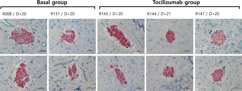
Figure 3.
Insulin and CD31 distribution in the islets transplanted to the liver of recipient monkeys. Liver samples taken at 20 or 21 days after pig islet transplantation was immunostained with anti-insulin and anti-CD31 antibodies. Results show well-engrafted islet β-cells (red) and lining of CD31+ endothelial cells (blue). In Basal group treated with basal immunosuppression, the transplanted islets had well-developed CD31+ cells and these endothelial cells were observed on the peripheral and intra islet areas. By contrast, in Tocilizumab group, intensity of insulin in β-cells and the extent of CD31+ cells in the intra-islet area were significantly lower than in Basal group.

