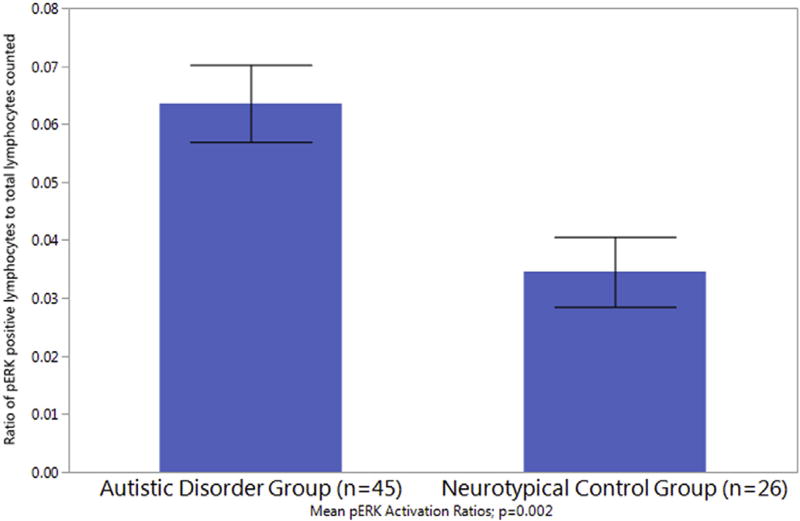
Fig. 1.
Lymphocyte Counting Phosphorylated ERK Analysis. Phosphorylated-ERK positive cells were identified and counted as activated based on the cytosolic translocation of p-ERK in the cell. The ratio of pERK positive lymphoctyes to total lymphocytes counted is significantly increased in the autistic disorder group as compared to the control group.

