Figure 3.

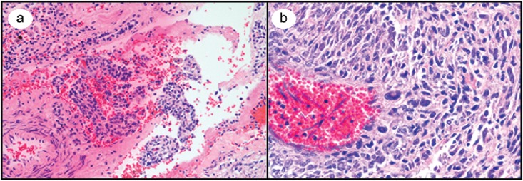
(a) Medium-power microscopic view of a hematoxylin and eosin–stained lung section containing a cyst with spindled and epithelioid cells. (b) High-magnification microscopic view of highly cellular kidney mass with multiple atypical cells.

(a) Medium-power microscopic view of a hematoxylin and eosin–stained lung section containing a cyst with spindled and epithelioid cells. (b) High-magnification microscopic view of highly cellular kidney mass with multiple atypical cells.