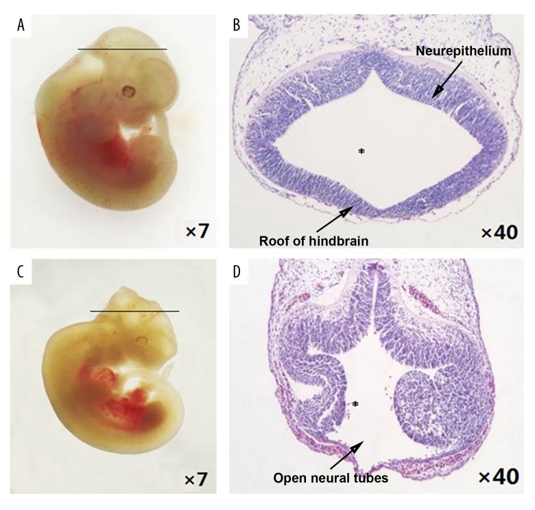
Figure 1.
Morphological changes in the neural tube defect (NTD) mouse model, gestational day (GD) 11.5. (A) Control embryos viewed under the dissecting microscope. (B) Photomicrograph of the light microscopy of the hematoxylin and eosin (H&E)-stained section of control mouse embryonic hindbrain. (C) NTD mouse model embryos viewed under the dissecting microscope. (D) Photomicrograph of the light microscopy of the hematoxylin and eosin (H&E)-stained section of the NTD mouse model embryos. Slice site: * fourth ventricle.

