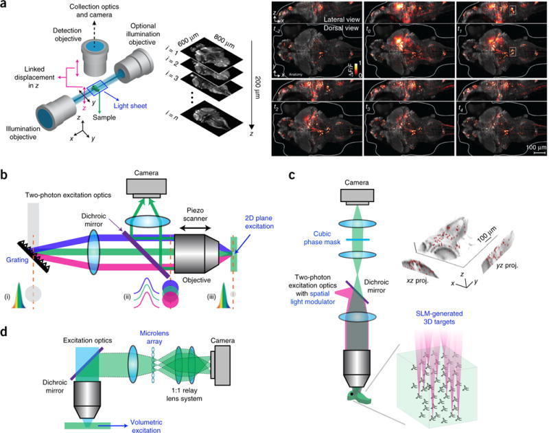
Figure 2.

Wide-field imaging. (a) Left, a schematic of a light-sheet microscope. Right, whole-brain neuronal activity of a larval zebrafish recorded with a light-sheet microscope. Brighter hues represent active neurons. Reprinted and adapted from ref. 3, Macmillan Publishers Limited. (b) Schematic of a microscope using temporal focusing. (i–iii) Temporal and spatial cross-section profiles of the laser beam impinging on the grating (i), at the back aperture (ii) and focal plane of the objective (iii) are shown. Colors indicate different spectrum components. Adapted from ref. 28, Macmillan Publishers Limited. (c) Holographic microscope with extended depth of field. Right, calcium imaging of 49 neurons targeted simultaneously on a zebrafish. Image reprinted from ref. 34, Frontiers. (d) Schematic of a light-field microscope. Adapted from ref. 37, Macmillan Publishers Limited.
