Figure 3.

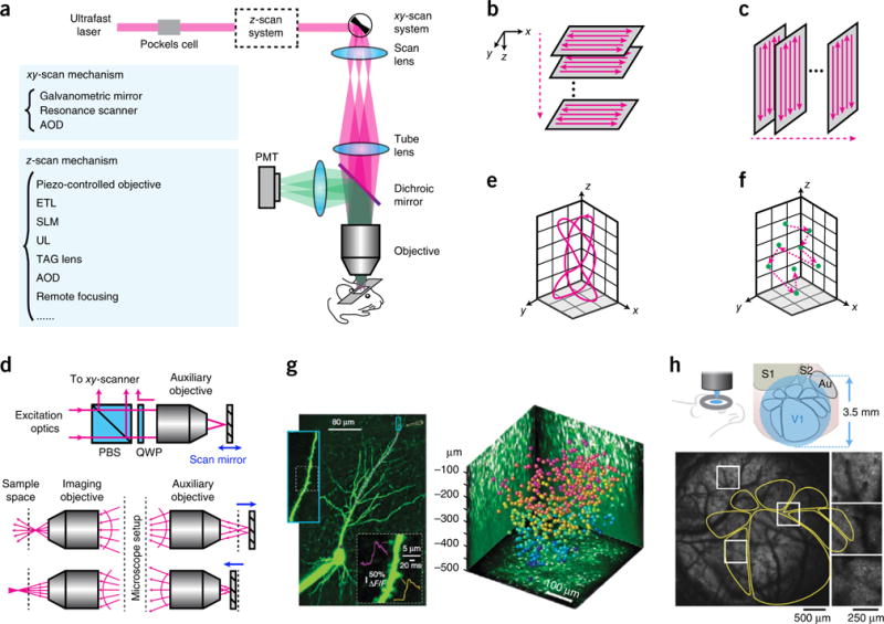
Two-photon microscopy. (a) Optical layout of a two-photon laser scanning microscope. (b) Traditional scan trajectory for volumetric imaging. (c) Scan trajectory enabled by ultrasound or TAG lens. (d) Principle of remote focusing. PBS, polarization beam splitter. QWP, quarter waveplate. Adapted from ref. 48 with permission from the National Academy of Sciences. (e) Custom scanning. Adapted from ref. 48 with permission from the National Academy of Sciences. (f) Random-access scanning in a 3D volume. (g) Examples of data taken with 3D random-access hopping microscope using AODs. Left, calcium imaging of a CA1 pyramidal cell; purple dots represent the scanning points in a dendrite. Right, neurons color coded to depth in mouse visual cortex, imaged with AODs. Reprinted from ref. 53, Macmillan Publishers Limited. (h) Large FOV microscope. Bottom, calcium imaging of a transgenic mouse expressing GCaMP6s. The yellow outlines indicate the anatomy mapped out by intrinsic signal optical imaging. Reprinted from ref. 57, Macmillan Publishers Limited.
