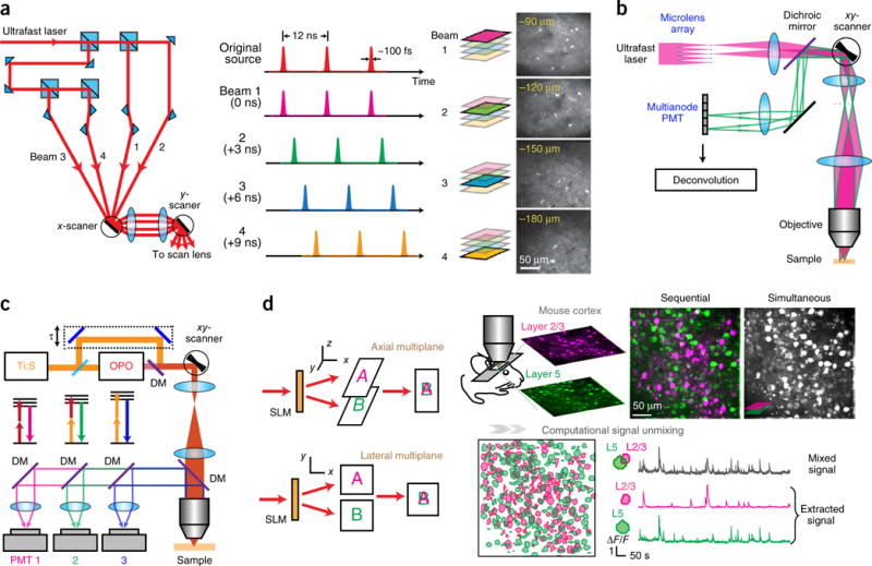
Figure 4.

Multiplexing two-photon microscopy. (a) Temporal multiplexing. The single laser beam is split, and one beamlet is delayed such that the pulse trains are interleaved in time. Right, example of a four-plane calcium imaging on mouse brain. Reprinted from ref. 58, Macmillan Publishers Limited. (b) Multifocal multiphoton microscope. Two beamlets are ray-traced. Adapted from ref. 59 with permission from the Optical Society of America. (c) Wavelength multiplexing. Pulse trains from two lasers (Ti:S, Ti:Sapphire and OPO) target two fluorophores with different two-photon absorption spectra. DM, dichroic mirror. Adapted from ref. 62, Macmillan Publishers Limited. (d) Holographic simultaneous multiplane imaging. An SLM splits the laser beam to illuminate different axial planes simultaneously. The right panel shows simultaneous calcium imaging on layers 2/3 and 5 on a GCaMP6f-transfected mouse visual cortex in vivo and post hoc signal separation. Reprinted and adapted from ref. 44 with permission from Elsevier.
