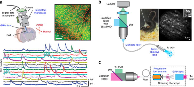
Figure 6.

Imaging freely behaving animals. (a) Epifluorescence miniature microscope. Top right, mean fluorescence image from a head-mounted microscope in a behaving GCaMP3-transfected mouse. Identified cells are shown in red. Bottom, fluorescence signals for 15 cells. Reprinted and adapted from ref. 89, Macmillan Publishers Limited. (b) Epifluorescence fiberscope. Right, a freely behaving mouse with the fiberscope fixed to its skull and calcium imaging of molecular layer interneurons recorded with structured illumination. Inset, single interneuron with a portion of a dendrite (arrow). DM, dichroic mirror. Reprinted and adapted from ref. 91 with permission from Elsevier. (c) Two-photon scanning fiberscope. Adapted from ref. 94 with permission from the Optical Society of America.
